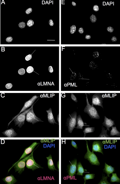
FIGURE 5.
MLIP co-localizes with lamin A/C and PML bodies in mouse C2C12 myoblasts. A–D, mouse C2C12 myoblasts were analyzed by indirect immunofluorescence microscopy (Carl Zeiss AxioImager Z1 Microscope) with antibodies against MLIP and lamin A/C. DNA was stained with DAPI (A), lamin A/C (B), and MLIP (C). D, merged images of DAPI (blue), lamin A/C (red), and MLIP (green) staining from A–C. Arrow indicates nuclear envelope and co-localization of MLIP with lamin A/C. E–H, C2C12 myoblasts were analyzed by indirect immunofluorescence and sequential scanning confocal microscopy with antibodies against MLIP and PML. DNA was stained with DAPI (E), PML (F), and MLIP (G). The arrows indicate co-localization of MLIP with PML. H, merged images of DAPI (blue), PML (red), and MLIP (green) staining from E–G. Scale bar, 10 μm.

