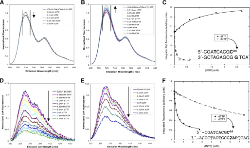
FIGURE 4.
Solution fluorescence studies of BF bound to double-stranded DNA containing a dideoxy-terminated primer strand. A–C, fluorescence resonance energy transfer between a fluorescent donor attached to the n-6 position of the DNA primer strand (green) and a Cy3 acceptor attached to Cys691 in the finger domain of BF wild type. Donor emission decreases upon titration with the complementary dCTP (A) but increases upon titration with a dTTP mismatch (B) as indicated by arrows. Emission intensities were integrated over the 511–530-nm interval (black vertical lines) and fit to a single-site binding isotherm (C) with Kd values of 29 mm for dCTP and 1.0 mm for dTTP. The DNA sequence is shown in the inset. D–F, change in emission intensity of 2-aminopurine placed in the template acceptor base position (magenta) upon titration of BF wild type with the complementary dTTP (D) and a dCTP mismatch (E) were integrated over the 330–460-nm interval and fit to single-site binding isotherms with Kd,dTTP = 0.29 mm and Kd,dCTP = 11 mm for BF wild type.

