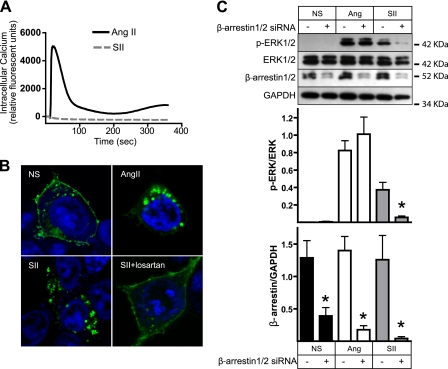
FIGURE 1.
Distinct efficacy of SII and AngII in HEK-AT1AR cells. A, effect of SII and AngII on intracellular calcium flux. Serum-deprived HEK-AT1AR cells were stimulated with SII or AngII, and time-dependent changes in intracellular calcium were measured using a fluorescence imaging plate reader. Representative traces are shown from one of three separate experiments that produced identical results. B, effect of SII and AngII on internalization of GFP-tagged AT1AR. Serum-deprived HEK cells transiently expressing AT1AR-GFP were treated for 30 min with vehicle (NS), AngII, or SII prior to fixation. Losartan (10 μm) was added to some dishes 10 min before SII stimulation. The cellular distribution of GFP was visualized by confocal fluorescence microscopy. Representative fields are shown from one of three separate experiments that produced identical results. C, effect of down-regulating β-arrestin expression on SII- and AngII-stimulated ERK1/2 phosphorylation. HEK-AT1AR cells were transfected with β-arrestin 1/2 targeted or control scrambled siRNA prior to serum starvation and stimulation for 5 min with SII or AngII. The upper panel depicts representative immunoblots of phospho- and total ERK1/2, β-arrestin 1/2, and GAPDH from one of three experiments. The upper bar graph depicts the mean ± S.E. of phospho-ERK1/2 normalized to the total ERK1/2. The lower bar graph depicts the mean ± S.E. of β-arrestin 1/2 expression normalized to GADPH. *, t test p < 0.05, less than cells treated with control siRNA (n = 3).

