FIGURE 4.
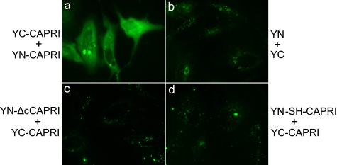
Detection of CAPRI dimerization in living cells using BiFC. To detect CAPRI dimers in living cells, CAPRI constructs were fused N-terminally to either the N-terminal (YN) or the C-terminal (YC) fragment of YFP protein, and HeLa cells were transfected to coexpress the following: either YN-CAPRI and YC-CAPRI (panel a) or YN and YC only (panel b); YC-CAPRI with a YN-mutant of CAPRI that lacks the 41 C-terminal AA (YN-ΔcCAPRI) (panel c) or YC-CAPRI with YN-mutant L775S/L778H of CAPRI (YN-SH-CAPRI) (panel d). Fluorescence signals were only detectable when CAPRI dimerization brought both halves of YFP into sufficiently close proximity to reconstitute the mature YFP fluorophore. In each group, over 200 cells were examined. Fluorescence signals were absent in the cells transfected with the constructs of panels b–d, respectively. 77% of cells transfected with the constructs of panel a were fluorescence-positive. The photographs shown are representative of three independent experiments. Scale bars, 20 μm.

