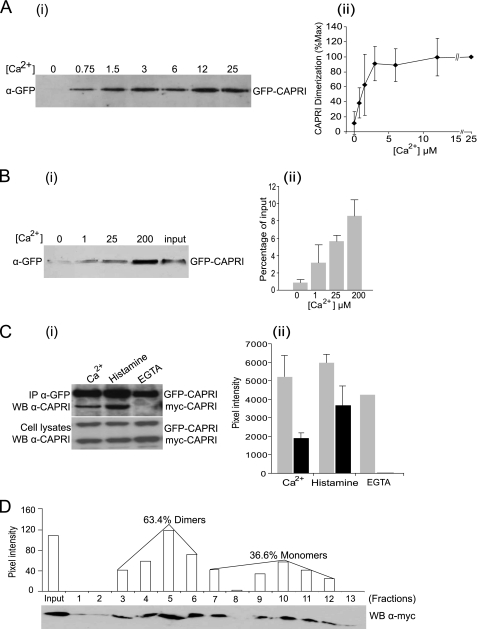
FIGURE 5.
Ca2+ dependence of CAPRI dimerization. A, CAPRI dimerization requires the presence of Ca2+. GFP-CAPRI was overexpressed in COS-7 cells. The cells were lysed in buffer containing 2 mm Ca2+. Immunoprecipitation was carried out with anti-GFP polyclonal antibody and protein A-Sepharose beads, and the beads were washed in buffers containing the indicated concentrations of Ca2+. Dimerized GFP-CAPRI was eluted with lysis buffer containing 1 mm EGTA and subjected to Western blotting with anti-GFP monoclonal antibody. Panel i, Representative Western blot. Panel ii, quantification by densitometric scanning of Western blots. Data are mean ± S.D. of three independent experiments. B, Ca2+ is sufficient to drive dimerization of purified GFP-CAPRI in vitro. Monomeric GFP-CAPRI was prepared as detailed under “Experimental Procedures,” and one-half was kept in solution, and the other was immobilized on beads. Aliquots of immobilized and free GFP-CAPRI were recombined in the presence of the indicated concentrations of Ca2+. Dimerized GFP-CAPRI was eluted with buffer containing 1 mm EGTA and subjected to Western blotting with anti-GFP monoclonal antibody. Panel i, representative Western blot. Input, 5% of the total GFP-CAPRI in each reaction. Panel ii, quantification of dimerized GFP-CAPRI as the percentage of total input. Data are mean ± range of two independent experiments. C, dimerization of CAPRI can be induced by histamine stimulation of HeLa cells. GFP-CAPRI and myc-CAPRI were co-overexpressed in HeLa cells. The cells were either lysed in buffer containing 1 mm Ca2+ or stimulated with 100 μm histamine before lysis in Ca2+ buffer or lysed in Ca2+-free buffer containing 0.5 mm EGTA, as indicated. Immunoprecipitation was carried out with anti-GFP monoclonal antibody, and the blots were probed with anti-CAPRI antibody. Panel i, representative Western blot (WB). Panel ii, quantification by densitometric scanning. Data are mean ± range of two independent experiments, except for EGTA, which are from one experiment. D, quantification of CAPRI monomers and dimers by gel filtration. myc-CAPRI was overexpressed in Cos-7 cells, and the cells were lysed in buffer with 500 μm Ca2+. The lysates were subjected to FPLC Superose 12 gel filtration column as detailed under “Experimental Procedures.” Column fraction aliquots (30 μl/fraction) were subjected to SDS-PAGE and Western blotting with anti-Myc monoclonal antibody. Quantification of the monomeric and dimeric forms of CAPRI was obtained by densitometric scanning. One-tenth input proteins were loaded in the input lane. The standard protein markers are blue dextran 2000 kDa, apoferritin 443 kDa, β-amylase 200 kDa, alcohol dehydrogenase 150 kDa, BSA 66.2 kDa, carbonic anhydrase 29 kDa, cytochrome c 12.4 kDa, and aprotinin 6500 Da.

