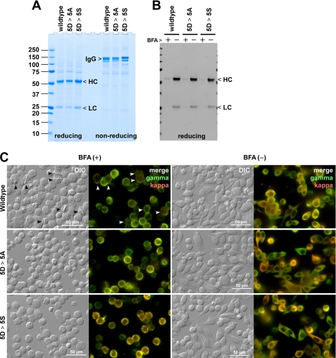
FIGURE 10.
Transiently transfected HEK293 cells develop intracellular crystals when ER export is blocked by brefeldin A. A, HEK293 cells were transiently transfected with IgG-encoding expression vectors. On day 6 post-transfection, cell culture media were harvested, and the protein quality was analyzed by SDS-PAGE under reducing and non-reducing conditions. A sample equivalent to 5 μl of neat culture medium was loaded per lane. The expression titer was 75 mg/liter for wild type IgG, 66 mg/liter for the 5D-to-5A mutant (5D > 5A), and 81 mg/liter for the 5D-to-5S mutant (5D > 5S). B, BFA treatment blocks IgG secretion. Transfected cells were cultured in a fresh medium from day 2 to day 3 post-transfection for 24 h either in the presence or absence of 15 μg/ml brefeldin A. The amount of IgG secreted to the culture medium during the 24-h period was examined by Western blotting after resolving the proteins by SDS-PAGE under reducing conditions. C, on day 2 post-transfection, the cells were seeded onto glass coverslips and cultured statically for 24 h in the presence (left two columns) or the absence (right two columns) of 15 μg/ml BFA. On day 3 post-transfection, the cells were fixed with paraformaldehyde, permeabilized, and immunostained with FITC-conjugated goat anti-γ chain and Texas Red-conjugated goat ant-κ chain antibodies. The wild type IgG-expressing cells that developed detectable intracellular crystals are marked by arrowheads. The “merge” is a digital overlay of anti-γ staining (green) and anti-κ staining (red). Crystals developed only in cells expressing wild type IgG under BFA treatment. Scale bars, 50 μm.

