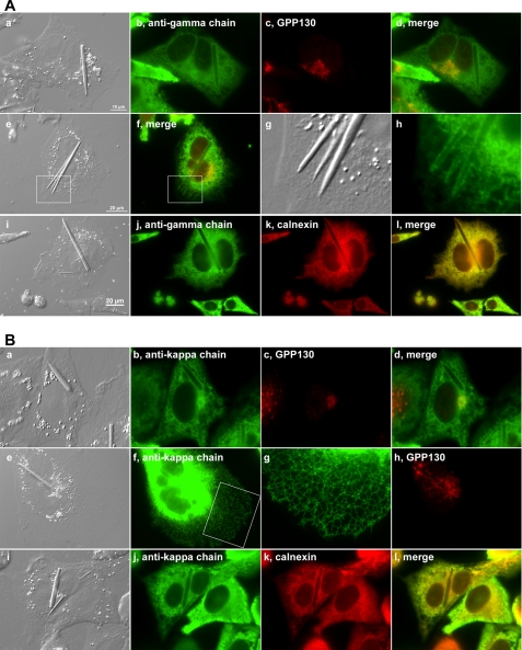
FIGURE 4.
Model IgG accumulates in ER. Suspension-cultured cells were allowed to spread on polylysine-coated glass coverslips for 24 h before cells were fixed with paraformaldehyde. A, panels a–l, cells were stained with an anti-γ chain antibody and co-stained with an anti-GPP130 or an anti-calnexin antibody. The white boxed area in panel i is digitally cropped and enlarged in panels k and l. B, panels a–l, cells were co-stained with anti-κ chain and anti-GPP130 antibodies. The white boxed area in panel f is digitally enlarged in panel g.

