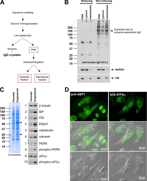
FIGURE 7.
Soluble milieu of ER contains various IgG folding intermediates. A, a schematic of homogenization procedures used in this fractionation study. B, after the fractionation, samples were resolved by SDS-PAGE both under reducing and non-reducing conditions. Western blotting was performed by using anti-human IgG (HC + LC) (top panel), anti-GAPDH (middle panel), and anti-transferrin receptor (TfR) (bottom panel). C, a whole cell extract was prepared from parental CHO cells and the engineered CHO cells expressing model human IgG. The lysate representing 6 × 10⁴ cells was loaded in each lane. Left panel, Coomassie staining of the whole cell lysates resolved under reducing conditions. Right set of panels, 10 selected proteins were detected by Western blotting. The names of the specific proteins are shown next to each blot. D, immunofluorescent staining of IgG-expressing engineered CHO cells with anti-XBP1 (left set of panels) or with anti-ATF6α (right set of panels). Scale bars, 20 μm. PDI, protein-disulfide isomerase.

