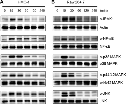
FIGURE 5.
spIL-33 induced the phosphorylation of IRAK1, NF-κB, p38 MAPK, p44/42 MAPK, and JNK. Human mast HMC-1 cells (A) and mouse macrophage Raw 264.7 cells (B) were treated with 50 ng/ml human spIL-33 as indicated time points. The phosphorylation of IRAK1, NF-κB, p38 MAPK, p44/42 MAPK, and JNK was significantly increased at 15 min and then drastically decreased at 60 min. However, phospho-NF-κB remained until 240 min in HMC-1 cell. The bottom of each panel exhibits the expression level of nonphosphorylated signaling molecule in cell lysates to show an equal amount of protein sample was loaded in each lane. The data represent one of three independent experiments.

