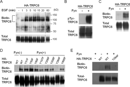
FIGURE 1:
Phosphorylation of TRPC6 Y284 is necessary for its trafficking to the plasma membrane. (A) Induction of membrane trafficking of TRPC6 by EGF. HEK293T cells expressing HA-TRPC6 were stimulated by EGF (200 ng/ml) for the indicated times. The cells were surface biotinylated with Sulfo-NHS-SS-Biotin, and the streptavidin-agarose–bound proteins were analyzed by Western blotting with α-TRPC6 antibody. The positions of the molecular-weight-marker proteins in kilodaltons are shown on the right side of the panel. A portion of each lysate (2%) was examined by Western blot to confirm the expression level of TRPC6 (bottom panel). (B) HA-TRPC6 expressed in HEK293T cells with or without constitutively active Fyn was immunoprecipitated and probed with anti-phosphotyrosine antibody. (C) HEK293T cells expressing HA-TRPC6 with or without Fyn were processed as in A. (D and E) Fyn and each of a series of phenylalanine substitution mutants of HA-TRPC6 were expressed in HEK293T cells (D) or in cultured podocytes (E), and the surface biotinylation assay for TRPC6 was performed as in A. Representative data from three to five independent experiments are shown.

