FIGURE 3:
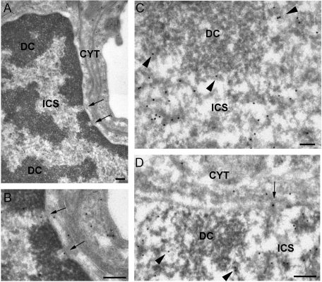
IEM localization of CBF-A in the cell nucleus. Thin sections of adult mouse brain were immunostained with the anti–CBF-A antibody SAK22. (A) Overview of a nucleus from a pericyte found wrapped around precapillary arterioles showing the typical appearance of dense chromatin regions (DC) and interchromatin space (ICS). The arrows point to two distinct nuclear pores. CYT, cytoplasm. (B) The same nuclear pores as in (A) at higher magnification. Note the presence of anti–CBF-A labeling in the central regions of the pores (arrows). (C and D) Examples of nuclear labeling in hippocampal neurons. The anti–CBF-A labeling is located in the interchromatin space (ICS) and in the perichromatin regions (arrowheads) but is excluded from the dense chromatin (DC). Note the labeling associated with a nuclear pore (arrow in D). The magnification bars represent 200 nm in A, B, and D and 100 nm in C.

