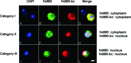
FIGURE 3:
Differential localizations of Ndt80 and Ndt80-bc. NDT80-HA/NDT80-bc-myc cells were stained with DAPI (A, E, and I), anti-HA antibodies (B, F, and J), and anti-myc antibodies (C, G, and K), to detect nuclei, Ndt80, and Ndt80-bc, respectively. Merged images are shown in (D), (H), and (L). Three categories of differential localization of Ndt80 and Ndt80-bc (A–D, E–H, and I-L) were as indicated and described in Results. Scale bar, 2 μm.

