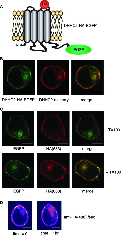
FIGURE 3:
Integration of DHHC2 into the plasma membrane and evidence of recycling. (A) Illustration of the predicted membrane topology of DHHC2 at the plasma membrane, highlighting the position of the inserted HA epitope and EGFP fluorescent tag. (B) Comparison of the intracellular localization of DHHC2-HA-EGFP and DHHC2-mCherry constructs cotransfected into PC12 cells. (C) PC12 cells transfected with the DHHC2-HA-EGFP construct were fixed and incubated in the absence (− TX100) or presence (+ TX100) of Triton X-100 for 6 min. The cells were then incubated with a mouse monoclonal HA antibody and subsequently with anti-mouse(633). (D) PC12 cells expressing DHHC2-HA-mCherry were incubated with anti-HA conjugated to Alexa Fluor 488 for 30 min at room temperature. The cells were then imaged directly, incubated on the microscope stage at 37°C for a further 60 min, and imaged again using identical settings. Scale bars on all figure panels represent 5 μm.

