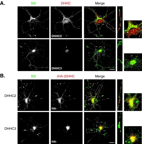
FIGURE 3:
Relative subcellular distribution of stathmin 2 and endogenous DHHC2 and -3 in neurons. (A) Hippocampal neurons at 6 DIV were colabeled for stathmin 2 (St2, green) and DHHC2 or -3 (red). DHHC2 displayed a vesicle-like staining in the cell body and along neurites partially overlapping but not significantly colocalized with the Golgi and vesicle-like labeling of stathmin 2. The endogenous DHHC3 staining was concentrated at the Golgi where it displayed a significant colocalization with stathmin 2 labeling. (B) Hippocampal neurons transfected at 6 DIV with HA-DHHC2 or -3 were colabeled for endogenous stathmin 2 (St2, green) and for exogenous DHHCs (red) with anti-HA antibodies. Contrary to DHHC2, DHHC3 is a Golgi-restricted DHHC PAT that colocalized with stathmin 2 at the Golgi. DHHC2 staining showed a punctuate labeling within the soma and neurites that colocalized partially with the ones of stathmin 2. Bars = 10 μM; boxed areas in the merged images are shown at higher magnification.

