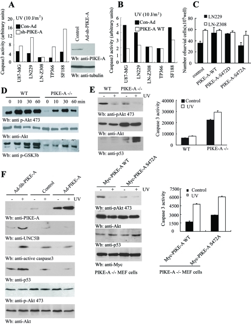
FIGURE 5:
PIKE-A regulates DNA damage-mediated apoptosis in glioblastoma cells. (A) Depletion of PIKE-A makes glioblastoma cells vulnerable to UV-triggered apoptosis. Activation of caspase-3 was quantified in glioblastoma cells infected with control and sh-PIKE-A adenovirus. Control and sh-PIKE-A adenovirus were used to infect different glioblastoma cells. Forty-eight hours after infection, the cells were exposed to 10 J/m2 of UV. Quantitative apoptotic assay in various cell lines is shown. Immunoblotting analysis of PIKE-A knockdown in LN-Z308 cells (right). (B) PIKE-A overexpression represses UV-triggered apoptosis. PIKE-A infected U87-MG, LN229, and SF188 cells revealed the decreased caspase-3 activity, but LN-Z308 and TP366 cells remained the same compared to control. (C) Akt phosphorylation of PIKE-A is required for its prosurvival activity in LN229 cells. LN229 and LN-Z308 glioblastoma cells were transfected with control and various PIKE constructs, followed by UV stimulation. Apoptosis was analyzed after 24 h. (D) PIKE-A is required for FBS-stimulated Akt activation. Wild-type and PIKE-A −/− MEF cells were treated with 10% FBS at various time points. The cell lysates were analyzed with anti–p-Akt 473, anti-Akt, and anti–p-GSK3 antibodies, respectively. (E) Wild-type but not PIKE-A Ser-472A mutant prevents PIKE-null MEF cells from UV-induced apoptosis. Top, wild-type and PIKE-A–null MEF cells were stimulated with UV. After 24 h, the cells were analyzed by immunoblotting with various indicated antibodies and caspase-3 activity was quantified by Caspase-Glo 3/7 assay. Bottom, PIKE-A–null MEF cells were transfected with Myc-PIKE-A WT or Ser-472A followed by UV stimulation. Akt activity and p53 levels were analyzed (bottom left). Caspase-3 activity was quantified by Caspase-Glo 3/7 assay (bottom right). (F) Knocking down of PIKE-A up-regulates p53 and UNC5B, enhancing apoptosis. TP366 cells were infected with control adenovirus or adenovirus expressing shRNA or wild-type PIKE-A. Over 24 h, the infected cells were exposed with UV. After 24 h, the cell lysates were analyzed with various antibodies as indicated.

