
Fig.2.
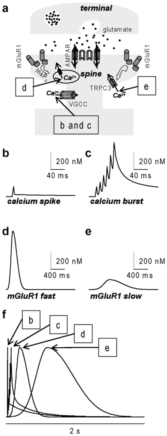
Schematic of four [Ca2+]i signals associated with excitatory synaptic transmission. (a) Associated with glutamate release from presynaptic terminals and depolarisation due to AMPA receptor (AMPAR) activation, [Ca2+]i can elevate via Ca2+ entry through VGCCs (signals b and c), via Ca2+ release from stores triggered by mGluR1 activation and InsP3 (signal d) and via Ca2+ entry through TRPC3 triggered by mGluR1 activation (signal e). (b) [Ca2+]i signal associated with one calcium spike: peak ~100–200 nM, duration ~10 ms; estimate from [14]. (c) [Ca2+]i signal associated with a burst of 6 calcium spikes at 100 Hz (calcium bursts): peak 0.5-2 μM, duration ~80 ms; estimate from [14]. (d) [Ca2+]i signal mediated by mGluR1 and Ca2+ release from stores (fast mGluR1): peak: ~1 μM, delay from mGluR1 activation ~50 ms, duration ~100 ms; estimate from [26]. (e) [Ca2+]i signal mediated by mGluR1 and slow Ca2+ influx (slow mGluR1): peak: ~100–200 nM, time-to-peak from mGluR1 activation ~0.5-1s, duration ~1s; estimate from [26, 30]. (f) The four [Ca2+]i signals normalised in amplitude and superimposed.