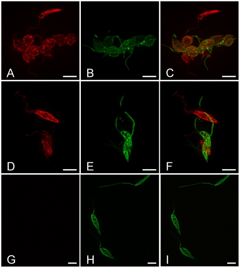
Figure 3. Hybrid promastigotes found in the sand fly midgut.
Images from the Olympus CellR 567 system showing dual fluorescence in Leishmania from a P. perniciosus female 2 days PBM (A–C). The female was infected with LEM 4265 (GFP transfected) and Gebre-1 (RFP transfected) parental strains. A, B, C, group of 10 hybrids together with four parasites showing red fluorescence only, D–F, group of two hybrids with two red parasites, G–I, GFP controls. A, D, G, images from red fluorescence, B, E, H, images from green fluorescence and C, F, I, merged images, scale bar: 5 µm.

