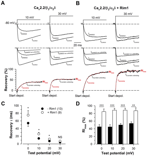
Figure 4. Rim1 promotes Cav2.2 channel recovery from G-protein inhibition.

A, Representative normalized Ba2+ current traces elicited at +10 mV and +30 mV before (IControl) and after 10 μM DAMGO application (IDAMGO) for Cav2.2/β3/α2δ1b channels (top panel). Corresponding traces showing the amount of current that is present in IDAMGO (IDAMGO wo unbinding) are also shown (middle panel) and used to calculate IG-protein unbinding, the time dependence of G-protein dissociation from the channel (bottom panel). IG-protein unbinding was fitted with a mono-exponential function (red dashed lines) in order to determine the time constant of recovery from G-protein inhibition (τrecovery) and the maximal extent of recovery (RImax). B, Legend as in (A) but for cells expressing Cav2.2/β3/α2δ1b/Rim1 channels. Corresponding mean values of τrecovery (C) and RImax (D) for Cav2.2/β3/α2δ1b (filled symbols) and Cav2.2/β3/α2δ1b/Rim1 channels (open symbols) as a function of membrane potential. RImax values were significantly increased in the presence of Rim1 for all potential values studied.
