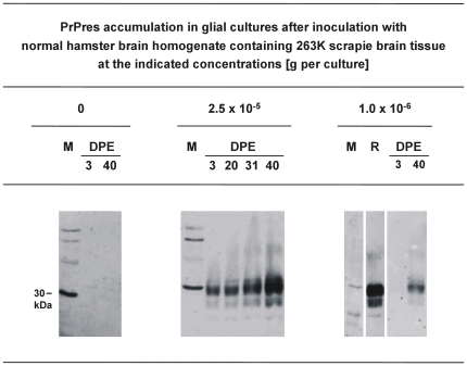
Figure 7. PrPres-accumulation in glial cell cultures exposed to scrapie brain homogenate.
Western blot detection of PrPres, the proteinase K–resistant core of misfolded PrP, at the indicated days post initial exposure (DPE) in glial cell cultures from hamsters inoculated with NBH containing 0, 2.5×10⁻⁵, or 1.0×10⁻⁶ g 263 K scrapie brain tissue. Lanes DPE represent 3.8 µl-aliquots from resuspended cell culture pellets harvested at the indicated days post initial exposure. Lane M, molecular mass marker; the prominent band (indicated by bar) corresponds to 30 kDa. Lane R, PrPres reference standard: PK-digested brain homogenate from scrapie hamsters corresponding to 5×10⁻⁷ g brain tissue.

