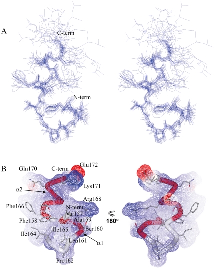
Figure 6. Overall structure of LC4 in SDS micelles at pH 4.5 by 1H-NMR.
(A) Stereoview illustrating a trace of the backbone atoms for the ensemble of the 20 lowest energy structures, showing the heavy atoms of the side chains (residues Val157-Leu174). The structures in the well-ordered region (residues Val157-Glu172) was superimposed over the backbone atoms. (B) Surface representation and ribbon diagram of LC4 showing the side chains (residues Val157-Glu172). The helical regions (α1 and α2) are shown in red.

