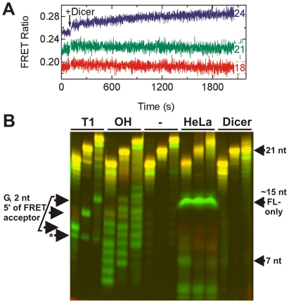
Figure 4. Processing of doubly-labeled dsRNAs of varying length by HeLa S100 cytosolic cell extract and purified human Dicer.
(A) Steady-state FRET time traces of 18-, 21-, and 24-nt dsRNAs as indicated upon Dicer addition; only the 24-nt dsRNA time trace slightly increases. (B) Denaturing FRET gel revealing the degradation patterns of the 18-, 21-, and 24-nt dsRNAs (from left to right in each triplet of bands) as a function of time, alongside RNase T1 (“T1”, leading to cleavage 3′ of G as indicated by arrows on the left) and alkali (“OH”) ladders. “-” indicates incubation in buffer only, while the last two triplets of bands show samples incubated in HeLa cell extract and purified Dicer enzyme, respectively. Arrows to the right mark specific degradation products.

