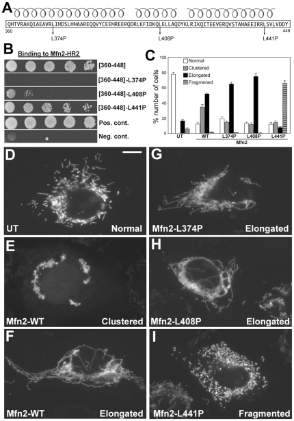
Figure 3. Differential binding effects of three HR1 helix-breaking mutation.
(A) Three leucine-to-proline mutations (L374P, L408P, and L441P) made in each of the three predicted helices in the Mfn2[360–448]. (B) Y2H assays testing interactions of three HR1 mutants with Mfn2-HR2. The Mfn2[360–448]-L374P and L408P disrupted the HR2 interaction whereas the L441P mutation maintained it. (C–H) Differential effects of three HR1 helix-breaking mutants on mitochondrial morphology. Clone 9 cells containing GFP-labeled mitochondria were transfected with Myc-tagged wild type Mfn2, Mfn2-L374P, Mfn2-L408P, or Mfn2-L441P, and mitochondrial morphology was evaluated in transfected cells that were identified by anti-Myc staining. (C) Cell counting for different mitochondrial morphologies among transfected cells. UT: untransfected. Error bars represent SEM. While normal tubular mitochondria were seen in untransfected cells (D), overexpression of wild-type Mfn2 induced mitochondrial clustering (E) as well as elongation of mitochondrial tubules (F). Mitochondria were overly elongated and entangled in the majority of cells overexpressing Mfn2-L374P (G) or Mfn2-L408P (H). Cells overexpressing Mfn2-L441P contained fragmented mitochondria (I). Scale bar: 10 µm.

