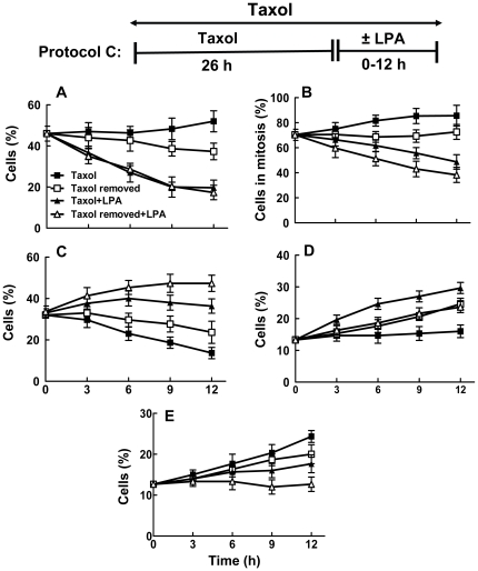
Figure 4. Lysophosphatidate releases MCF-7 cells from Taxol-induced arrest in G2/M and cell death.
MCF-7 cells were preincubated with 10% delipidated serum and 50 nM Taxol for 26 h. Taxol was then maintained or removed in the presence of 10% delipidated serum and 5 µM LPA was added as indicated (Protocol C). Cells were then stained with DAPI, anti-phospho-histone and with anti-tubulin at the times indicated. The Panels show the percentage of: A) cells in mitosis, B) percentage of cells in mitosis that had abnormal spindles, C) mononucleated cells, D) multinucleated cells and E) dead and dying cells. Total number of cells (100%) includes mononucleated, multinucleated, dead and dying and mitotic cells. Time zero shows 26 h pretreatment with Taxol. Results are means ± SD (where large enough to be shown) for three independent experiments.

