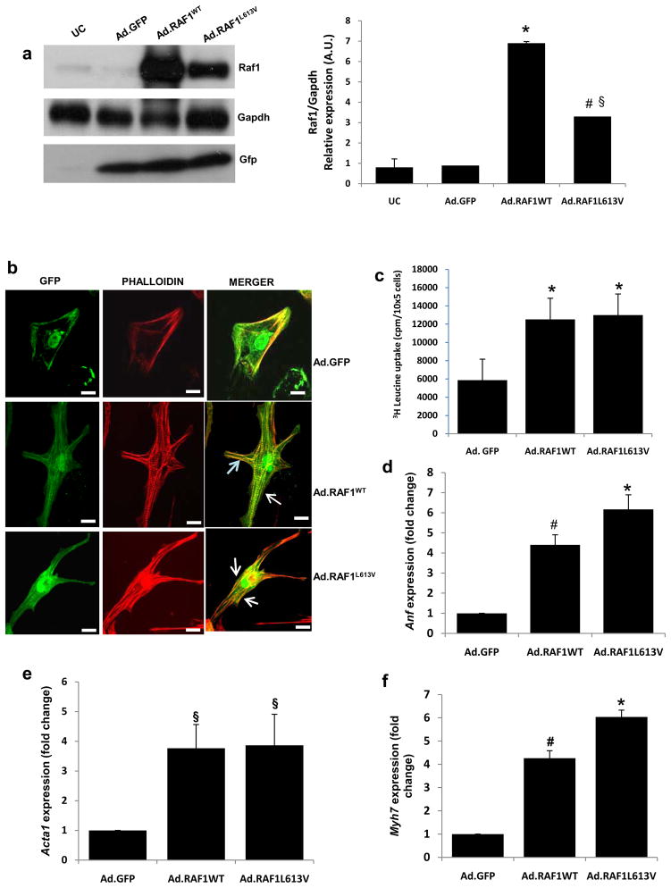
Figure 1. Overexpression of wild-type and L613V RAF1 proteins induces cardiomyocyte hypertrophy.
Neonatal rat cardiomyocytes were infected with adenoviruses encoding GFP alone (Ad.GFP), wild type RAF1 (Ad.RAF1WT) or a mutant RAF1 (Ad.RAF1L613V) and harvested after 72 hours. (a) Representative immunoblots with total lysates that were probed with anti-Raf1 (top panel), anti-Gapdh (middle panel) and anti-Gfp (lower panel) antibodies. Gapdh levels were used as loading control and Gfp levels were indicative of the efficiency of viral production. Uninfected cardiomyocytes (UC) were used as a negative control. Protein expression levels were normalized to respective Gapdh and expressed as relative expression. Data are mean values ± SD of three independent experiments §p <0.05 vs. Ad.GFP; *p <0.01 vs. Ad.GFP; #p <0.05 vs. WT (b) Cardiomyocytes infected with the viruses indicated at the right expressing GFP (left column) and stained with phalloidin to detect sarcomeric α-actinin(middle column) and merged (right column). Cells were imaged with confocal laser microscopy. Scale bar- 387×387nm. (c) [3H]-leucine incorporation in infected cardiomyocytes as an assessment of protein synthesis rates. *P <0.001 vs Ad.GFP. Data are mean values ± SD of three independent experiments. (d-f) Quantitative reverse transcription PCR analysis of fetal gene re-activation. Steady-state mRNA levels of (d) atrial natriuretic factor (Anf), (e) α-skeletal actin (Acta1) and (f) β-myosin heavy chain (Myh7) were performed. Expression levels were normalized to β-actin and expressed as fold change compared to level in the Ad.GFP cells. The mean fold induction ± SD of three independent experiments is shown. §p <0.05 vs. Ad.GFP; *p <0.001 vs. Ad.GFP; #p <0.01 vs. Ad.GFP.

