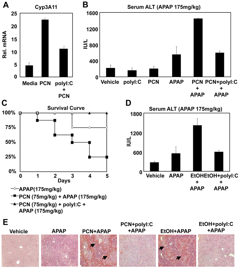
Figure 4. PolyI:C protects against APAP-mediated hepatotoxicity promoted by PCN or EtOH.
(A) Mice were treated with saline, PCN (75mg/kg, i.p.) or PCN and polyI:C (100µg, i.p.). After 24 hours, liver RNA was isolated and analyzed by Q-PCR. (B) Mice were treated with saline, polyI:C (100 µg, i.p.) or PCN (75mg/kg, i.p.) 24 hours prior to treatment with APAP (175 mg/kg, i.p.). (n=4, mean +/− SD, *P<0.05 compared to polyI:C untreated) (C) Mice (n=8) were treated with saline or polyI:C (100 µg, i.p.) and PCN (75 mg/kg, i.p.) 24 hours prior to administration of APAP (175 mg/kg, i.p.). Mice were followed for 5 days following treatment. (D) Alternatively, wild-type mice were given 20% EtOH ad libidum for 5 days, and saline or polyI:C were injected on Day 3 and Day 5. Mice were fasted 18 hours prior to APAP treatment (175 mg/kg) on Day 6. Six hours after APAP treatment, serum samples were isolated and analyzed for ALT levels. (E) Mice were treated as in B and D. Six hours post APAP treatment, mice were sacrificed and liver samples were isolated. Liver samples were formalin fixed and stained with H&E. Arrows indicate foci of necrosis.

