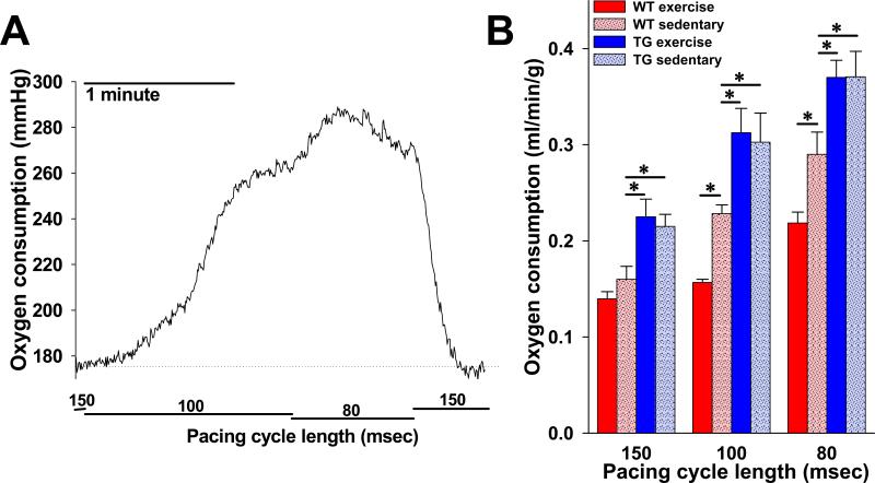
Figure 4. Rate of cardiac oxygen consumption in response to heart rate acceleration is reduced in animals with increased KATP channel expression following exercise.
A. Representative tracing of raw oxygen consumption data (perfusate oxygen tension – effluent oxygen tension) in response to pacing cycle length transitions in an isolated perfused heart from a WT sedentary animal. B. Summary statistics indicating steady state oxygen consumption rate (V̇O2), normalized to heart weight and coronary flow (see Methods), at each of three pacing cycle lengths in isolated perfused hearts from WT exercise (n=7), WT sedentary (n=7), TG exercise (n=4) and TG sedentary (n=5) mice (*p<.05 compared to WT sedentary). WT = wild-type, TG = Tg[αMHC-Kir6.1AAA].

