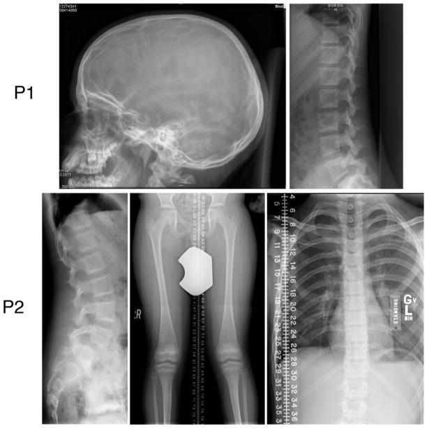
Figure 2.
Patient radiographs show a generalized decrease in bone mass. The skull radiograph of Patient 1 shows few wormian bones, while her vertebral bodies have os en os without compressions. Patient 2 also has os en os vertebral bodies, perhaps a reflection of prior bisphosphonate therapy, but without compressions. The lower extremities show genu valgum, gracile midshafts of the femora, dense traverse lines from cycled bisphosphonate therapy, tibial bowing and a healed left fibular fracture. His thorax is long and narrow.

