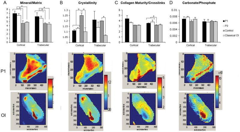
Figure 7.
Composition of iliac crest bone from patients with the C-propeptide cleavage site mutation, classical OI, and age-matched controls as determined by Fourier-Transform Infrared Imaging (FT-IR) Spectroscopy. The four parameters listed in the top row were measured for all cases and mean +/− SD for 3 areas of cortical and 3 areas of trabecular bone are shown, with * representing significant differences relative to the bracketed group, p<0.05 based on a t-test. Typical images from P1 and from a classic OI case are shown for each parameter. (A) Both C-propeptide mutation patients had an elevated mineral to matrix ratio in cortical and trabecular bone, compared to both two normal control and two classical OI samples. The mineral to matrix ratio as determined by FT-IR is directly related to the mineral content of the tissue. (B) Cortical crystallinity, a parameter related to the crystal size and perfection in the c-axis direction of the apatite crystals, was significantly decreased in the patients relative to controls and comparable to classic OI samples. (C) Cleavage site patients had an increase of trabecular collagen cross-links and a trend toward increased cortical cross-links. The collagen cross-link ratio is an indication of the maturity of the collagen, and does not provide information on the nature of the cross-links. (D) Carbonate to phosphate ratio (mineral composition), which indicates the extent of carbonate substitution in the apatites, did not vary significantly among samples.

