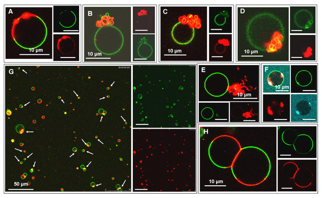
Figure 2.
All vesicles are phase separated CL-containing GUVs labelled by the NBD-PE (green) and Rh-PE (red) probes. Images A–E,G,H are GUVs interacting with Alexa Fluor 568 cyt c (red) and image F is a GUV in the presence of Alexa Fluor 633 labelled cyt c (blue). (A) Beading of the membrane can be observed in the Ld domain. (B–D) The Ld domains on the vesicles have collapsed into a folded structure where the folds of the membrane are resolvable under the confocal microscope. (E) The morphology of the Ld domain after cyt c-induced collapse appears to have a “wispy” morphology. (F) Two separate Ld domains coexisting in the same vesicle have collapsed into tight, folded structures. (G) Lower magnification image of many GUVs in the sample. White arrows indicate Ld domains that have collapsed to a folded structure after the addition of cyt c, thus demonstrating that the morphological transition we observe represents the majority behavior of vesicles in the sample. (H) Strong adhesion between the negatively charged, CL-containing Ld domains induced by the polycationic cyt c results in membrane deformation and a large osculating area between the two GUVs. The insets represent the separate fluorescence channels that are collected and superimposed to create the composite image.

