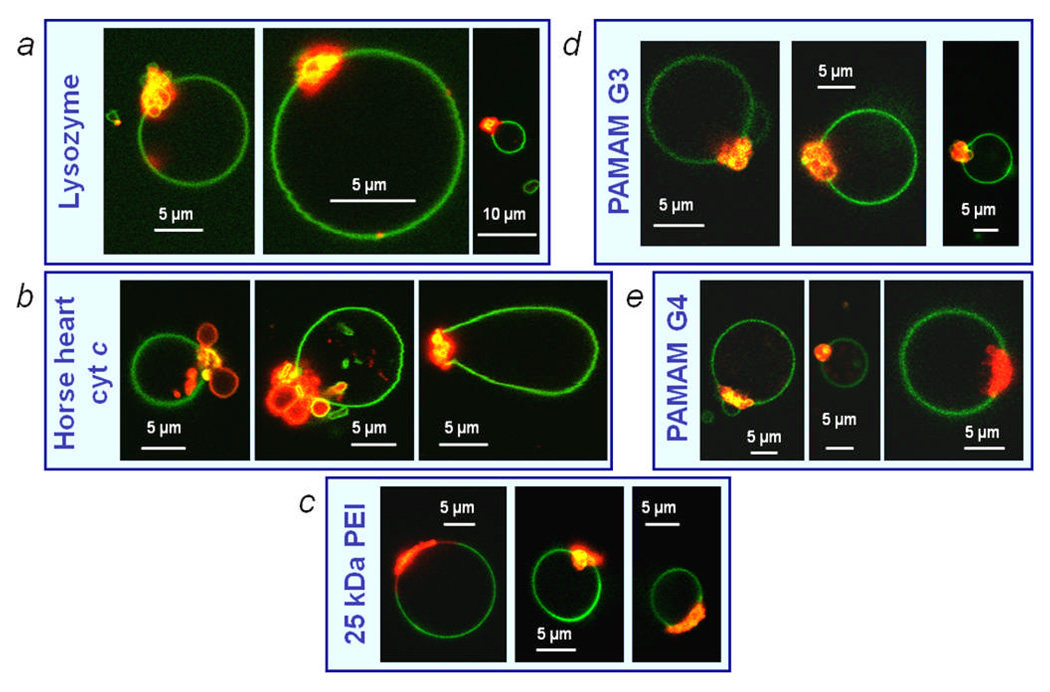
Figure 4.
Resultant vesicle morphologies of phase separated, CL-containing GUVs after incubation in solution with (a) lysozyme, (b) horse heart cyt c, (c) 25 kDa branched PEI, (d) generation 3 PAMAM dendrimer and (e) generation 4 PAMAM dendrimer. Rh-DPPE (red) labels Ld domains and NBD-PE (green) labels the Lo domains.

