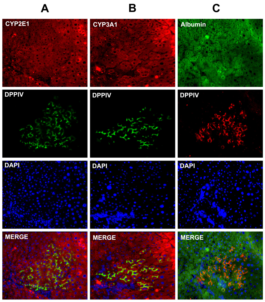
Figure 7. Naïve rAECs integrate and form clusters of mature hepatocytes upon transplantation into syngenic rats.
Immunofluorescence staining of serial frozen section of rat livers after transplantation of rAECs. Recipient animals were DPPIV− while transplanted rAECs were isolated from DPPIV+ tissues. Clusters of positive differentiated cells can be found into the host liver. (A) Double stain for DPPIV (green) and CYP2E1 (red). (B) Double stain for DPPIV (green) and CYP3A1 (red). (C) Double stain for DPPIV (red) and Albumin (green). Differentiated rAECs expressed hepatocyte markers at levels comparable to the surrounding liver.

