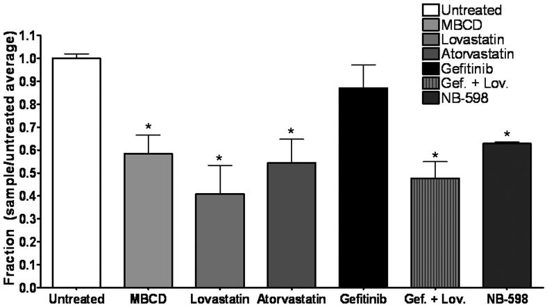
Figure 5. MBCD, lovastatin, atorvastatin, and NB-598 reduce cholesterol in breast cancer cells.
Fifty thousand cells were plated into 6-well plates and treated with 1.0 mM MBCD (1 h), 1.0 μM lovastatin (72 h), 1.0 μM atorvastatin (72 h), 1.0 μM NB-598 (72 h), 1 μM gefitinib (1 h), or a combination of 1.0 μM gefitinib (1 h) and 1.0 μM lovastatin (72 h) (Gef. + Lov.) in growth medium. Lysis was followed by protein quantification and cholesterol was measured using the Amplex Red cholesterol assay kit. Absorbance was converted to μg cholesterol/mL utilizing a cholesterol standard curve, and then samples were normalized to protein concentration for a final value in μg cholesterol/μg protein. Bars represent fraction of cholesterol with untreated samples as 1 (μg cholesterol/μg protein sample)/(μg cholesterol/μg protein untreated). Experiments were repeated at least three times in duplicate. Error bars represent the standard error of the mean. Statistical analyses were performed utilizing Student's t-test, * = p < 0.05 compared to untreated.

