
Figure 4.
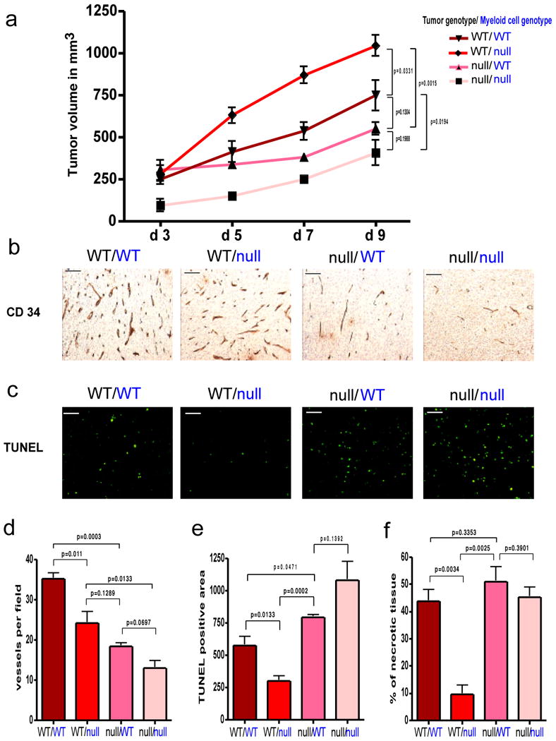
Effect of tumor cell-derived versus myeloid cell-derived VEGF on tumor angiogenesis and growth.
a, Growth curve analysis of wildtype (WT) and VEGF nullizygous (null) fibrosarcoma isografts (genotype labeled in black) implanted into WT-mice or Mut-mice (null) with a myeloid cell-specific deletion of VEGF (genotype labeled in blue) (n=4 for each group). b, CD 34 immunostaining on fibrosarcoma isografts. c, Detection of apoptotic cells in fibrosarcomas by TUNEL-staining. d, Quantitative analysis of CD 34-positive blood vessels (n=4). e, Quantification of TUNEL-positive cells (n=4). f, Assessment of tumor necrosis on fibrosarcoma midline sections. Shown is the perimeter of necrotic areas expressed as percentage of total tumor perimeter (n=4). Scale bar, 100 μm; error bars, s.e.m.