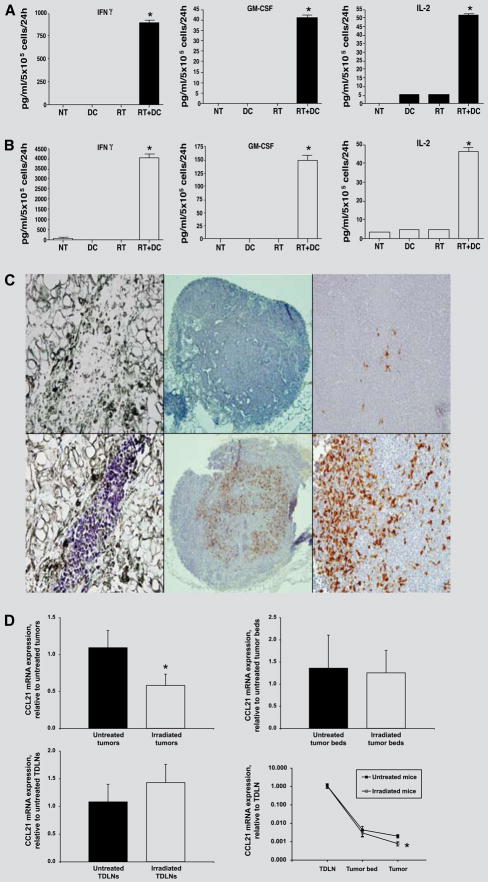
FIGURE 3.
A and B, Lymph node cells draining D5 tumors subjected to combined treatment with radiation plus DCs, but not to monotherapies, undergo effective in vivo priming to tumor antigens. Unpulsed-DCs were injected into irradiated D5 tumors. Control groups of mice received either no treatment (NT), DC only, or radiation only (RT). After 2 days, TDLN cells were harvested and analyzed for IFN-γ, GM-CSF, and IL-2 secretion in response to either D5 cells (A) or anti-CD3 mAb activation (B). Data are reported as the average concentration of cytokine (pg/ml) per 5 × 10⁵ responders per 24 h ± SE of triplicate samples. *, P<0.001 versus all other groups. C, Tumor irradiation enhances DC migration to the draining lymph node. CFSE-labeled unpulsed-DCs were injected into irradiated versus untreated s.c. D5 tumors either 1, 3, or 7 days after radiation. Tumors and TDLNs were harvested 24, 48, and 72 hours after each i.t. injection. Fluorescein-labeled cells were detected using an anti-FITC, horseradish peroxidase-conjugated antibody. Positive cells stained purple in tumor sections, and brown in lymph node sections. Representative fields from two independent experiments are shown [original magnification, X400 (left and right) or X100 (middle)]. Upper left, Tumor section stained with an isotype matched control antibody. Lower left, Tumor section stained with anti-FITC antibody. Upper middle and right, Section of a lymph node draining an untreated tumor. Lower middle and right, Section of a lymph node draining an irradiated tumor. D Tumor irradiation decreases CCL21 gene expression within the tumor mass by one-half compared with untreated tumors (upper left). But CCL21 gene expression within the tumor beds (upper right), and the tumor draining lymph nodes (lower left) does not change significantly between irradiated and untreated tumors. As a result, CCL21 concentration gradient between the tumor and the draining lymph node is increased (lower right). Expression of CCL21 mRNA within irradiated and untreated s.c. D5 tumors, tumor beds, and TDLNs was measured by quantitative real time RT-PCR. CCL21 mRNA expression was first normalized to the expression of HPGRT, and was then normalized to the average expression of CCL21 mRNA in untreated tumors (upper left), in untreated tumor beds (upper left), in untreated TDLNs (lower left) or in corresponding TDLNs (lower right). RNA was extracted from five tumors, three tumor beds, and three TDLNs per group, and gene expression was examined in individual samples. * p < 0.05. In upper right and lower left panels p is non-significant.

