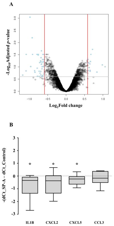
Figure 7.

Effects of SP-A treatment in the human amnion explants. A, Human reflected amnion explants obtained from TNL patients were treated with SP-A (1 μg/ml) for 12 h. A volcano plot showing the relationship between their fold changes (log2 of, x axis) and FDR-corrected p values (−log10 of, y axis). Positive values on the x-axis indicate genes whose expression increased after SP-A treatment, while negative values indicate genes down-regulated after SP-A treatment. B, Confirmative qRT-PCR data showing changes in the expression of select cytokines and chemokines. IL-1β, CXCL2, and CXCL5 mRNA expressions decreased significantly after treatment with SP-A (p < 0.05 for all comparisons). CCL3 mRNA expression was not significantly different. * p<0.05 by Wilcoxon signed rank test
