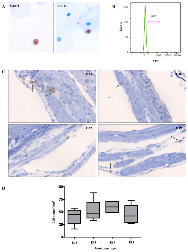
Figure 8.
Macrophages in the human amniotic fluid (A and B) and in the myometrium of C57BL/6 mice (C and D). A, Amniotic fluid cytologic specimens show CD68 immuno-positive macrophages among scattered epithelial cells. Case 4: a case of a patient presenting with intrauterine fetal demise at the gestational age of 21.4 weeks, Case 15: a case of a TNL patient at the gestational age of 38.7 weeks (original magnification X200; hematoxylin was used for counterstaining). B, A histogram from flow cytometric analysis of amniotic fluid cells does not show CD14 positive macrophages. The amniotic fluid was obtained from a TNL patient at the gestational age of 38 weeks. Green: CD14, Red: isotype. C, F4/80 immuno-positive macrophages (arrows) in the C57BL/6 myometrium obtained at various gestational ages: E13, E15, E17, and E19 original magnification X200; hematoxylin was used for counterstaining). D, The density of macrophages in the mouse myometrium did not change significantly across gestation. The numbers of F4/80 positive macrophages were counted in myometrial sections containing the mid-plane of the placenta from four pregnant mice at each gestational age (E13, E15, E17, and E19). Eight high-power fields corresponding to every 45° angle point starting from the mesometrial border in a clockwise direction, covering the entire myometrial circumference, were evaluated in each section. Image-Pro Plus 6.2 software (Media Cybernetics, Inc.) was used for the analysis.

