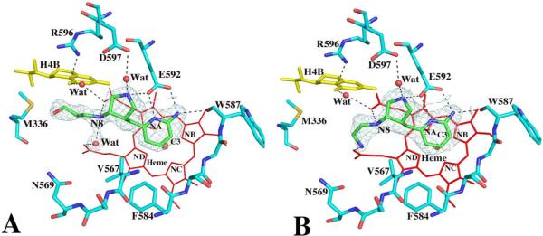
Fig. 2.
Active site structures of the wild type nNOS with inhibitor 4 (panel A) or 5 (panel B) bound viewed side by side in an identical orientation. Shown also the Fo – Fc omit map contoured at 3.0σ for each inhibitor. Hydrogen bonds are drawn with the dashed lines. The atomic color scheme for amino acids is: carbon, cyan or green; nitrogen, blue; oxygen, red; sulfur, yellow. The figures are made with PyMol (http:://pymol.sourceforge.net).

