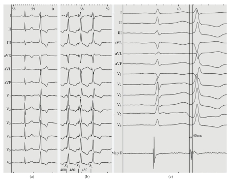
Figure 1.
(a) Baseline ECG showing a premature ventricular extrasystole displaying a qR pattern in lead V1; (b) intracardiac tracing during left ventricular mapping (MapD) showing the earliest local activation at the basal left ventricle; (c) pace mapping at this location demonstrated “perfect match” (12 of 12 leads) with the morphology of the premature ventricular extrasystoles.

