
Fig. 6.
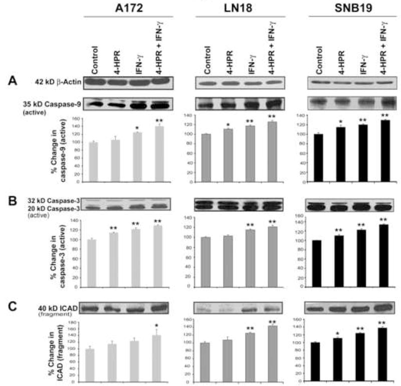
Western blotting for examination of activation of caspase-9 and caspase-3 and also formation of ICAD fragment in glioblastoma A172, LN18, and SNB19 cells. Treatments: control, 0.5 μM 4-HPR (5 days), 200 units/ml IFN-γ (2 dads), and 0.5 μM 4-HPR (3 days) + 200 units/ml IFN-γ (2 days). Representative Western blots (n ≥ 3) and bar graphs show changes in: (A) 35 kD active caspase-9, (B) 20 kD active caspase-3, and (C) 40 kD ICAD.