Figure 3.
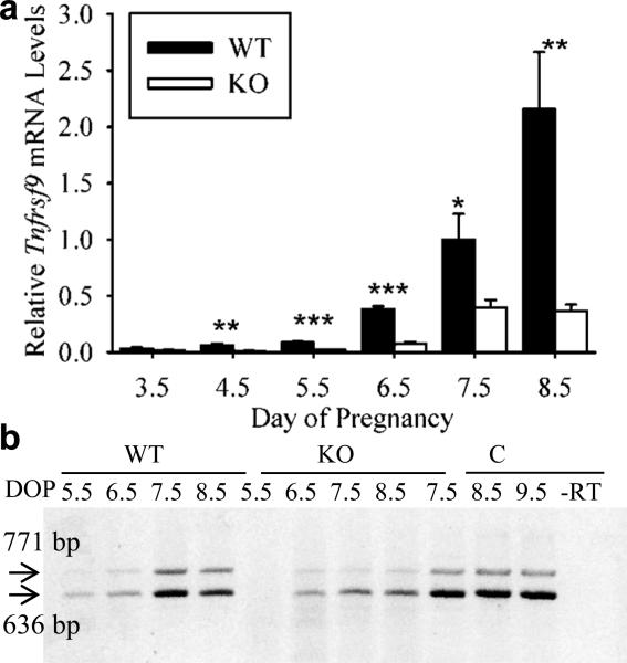
qRT-PCR and RT-PCR analysis of steady-state mRNA levels and presence of alternatively spliced mRNA forms of Tnfrsf9 in implantation site tissue segments of pregnant wild-type (WT) and Il15-knockout (KO) mice. (a) qRT-PCR analysis of relative mRNA levels of Tnfrsf9 mRNA in WT compared to KO IS tissue segments on Day 3.5-8.5 of pregnancy. Stars represent a significant difference in mRNA levels on a given Day of pregnancy between samples from WT and KO mice (*, P<0.05; **, P<0.005; ***, P<0.0005). (b) RT-PCR analysis of Tnfrsf9 splice variant expression implantation site (IS) tissue segments from the uteri of WT and KO mice on Day 5.5-8.5 of pregnancy and in conceptus tissues of WT mice from Days 7.5-9.5. RT-PCR results are representative of several (N=3) independent samples. Abbreviations: bp, base pair; DOP, day of pregnancy; –RT, no-reverse transcriptase control.

