Fig. 1.
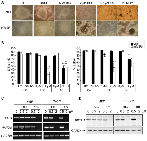
Treatment of hESCs with GSK-3 inhibitors induces differentiation. Shef-3 hESCs were treated with BIO, 1m or vehicle (DMSO) or left untreated (UT) and cultured for 7 days on either MEFs or Matrigel in mTeSR1 medium. (A) Images show the typical colonies that are formed. Scale bar: 1 mm. (B) hESCs were analysed by flow cytometry following immunostaining with antibodies against the pluripotency markers Tra-1-60 and SSEA4. Data show the mean percentage of positive cells (±s.e.m.) from at least three independent experiments. Statistical analysis was conducted using ANOVA and Dunnett's post hoc test to compare each treatment with the untreated control cells. *P<0.05; **P<0.01. An example of a histogram plot from a representative experiment is shown in supplementary material Fig. S2A. (C) RNA was extracted from the cells and RT-PCR analyses were performed using primers specific to the pluripotency genes OCT4 and NANOG and to the house-keeping β-actin-encoding gene. (D) Cell lysates (20 μg) were separated by SDS-PAGE and immunoblotting was performed using an antibody against OCT4. Blots were stripped and re-probed with anti-GAPDH antibodies to assess equal loading.

