Fig. 2.
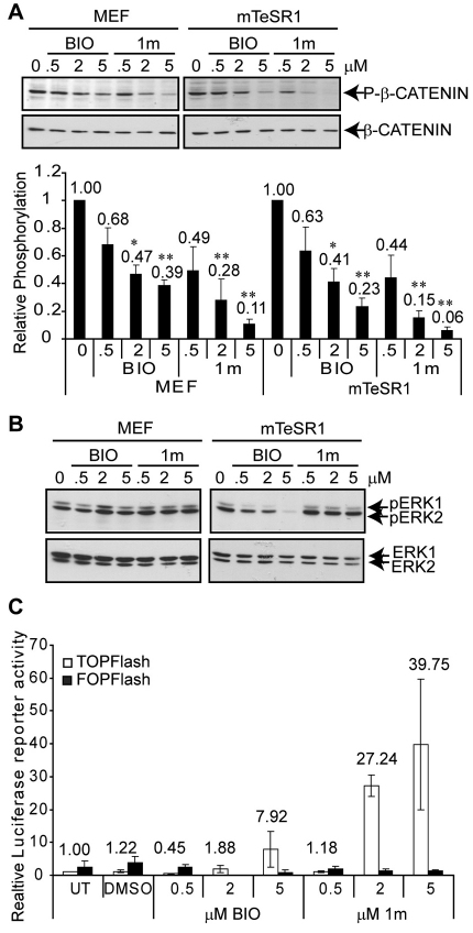
Treatment of hESCs with 1m inhibits GSK-3. (A,B) Shef-1 hESCs, cultured on MEFs or Matrigel in mTeSR1 medium, were treated with BIO or 1m for 30 minutes. Immunoblotting was performed to detect phosphorylated forms of β-catenin and ERK1/2. The same immunoblot in each case was re-probed for total β-catenin and ERK1 to assess loading. The bar graph shows the mean relative β-catenin phosphorylation levels (+s.e.m.; n=3). Data were analysed for statistical significance using two-tailed paired t-tests. *P<0.05; **P<0.01. (C) TOPFlash luciferase reporter assay following 24 hours of treatment of Shef-1 hESCs with BIO, 1m or vehicle (DMSO). Luciferase activity is expressed relative to the normalised TOPFlash luciferase activity in untreated (UT) control cells. A control reporter (FOPFlash), containing mutant TCF-binding sites, was also run in parallel. Data are means±s.e.m. for three independent experiments.

