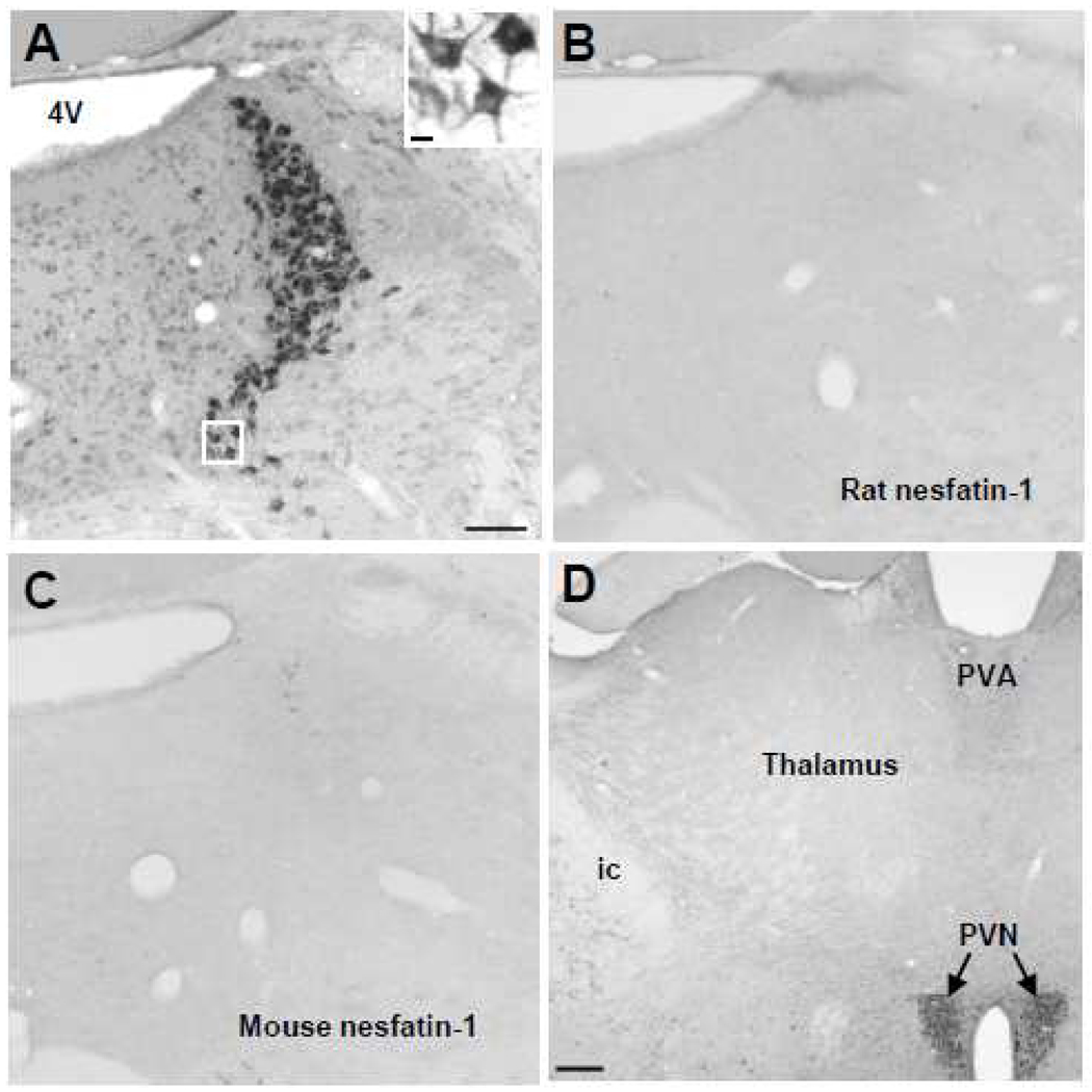
Fig. 2.
Specificity of the rat nesfatin-1 antibody shown by immunostaining and pre-absorption. Immunohistochemical staining of the locus coeruleus (LC) with the nesfatin-1 antibody (A, insert) and after preabsorption with either rat (B) or mouse (C) nesfatin-1 polypeptide. No immunostaining can be detected in the mouse LC after the rat nesfatin-1 antibody was pre-absorbed with rat or mice nesfatin-1 polypeptide. (D) shows site specificity with the lack of staining in most part of mouse thalamus by the nesfatin-1 antibody. Scale bar 100 µm in A is representative for B and C. The scale bar in the insert in A is 10 µm in C is 200 µm. Abbreviations: 4V, fourth brain ventricle; ic, internal capsule; PVA, anterior paraventricular thalamic nucleus; PVN, paraventricular nucleus of the hypothalamus.

