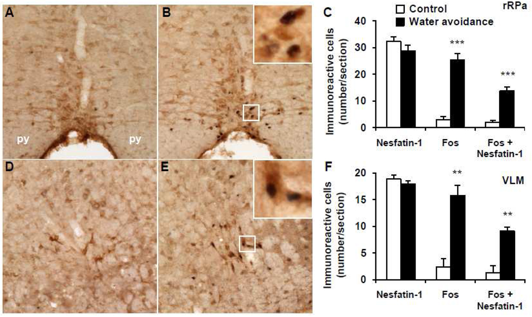
Fig. 6.
Representative double immunohistochemical staining for Fos (dark blue) and nesfatin-1 (brown) in the rostral raphe pallidus (rRPa) and ventrolateral medulla (VLM) in control mice (A, D) and after water avoidance stress (B, E). The inserts in B and E show higher magnification of neurons with Fos immunoreactivity co-localizing with nesfatin-1 immunoreactivity in the rRPa (insert B) and VLM (insert E). The scale bar in A is 100 µm representing the scale for all the panels and 10 µm in the inserts. Unilateral cell count/section in the rRPa (C) and VLM (F) as mean ± SEM of 4 mice/group. **p < 0.01, ***p < 0.001 compared with control. Other abbreviations: py: pyramidal tract.

