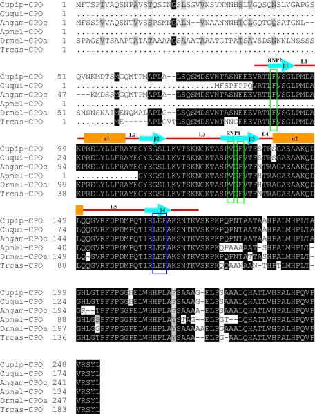
Figure 1.
Sequence alignment of deduced CPO amino acid sequences at the conserved C-terminal and a schematic diagram of the RNA recognition motif presented above the sequences. Sequence of Culex pipiens CPO (Cupip) was compared with CPOs from Culex quinquefasciatus (Cuqui), Anopheles gambiae (Angam), Apis mellifica (Apmel), Drosophila melanogaster (Drmel), and Tribolium castaneum (Trcas). Conserved amino acid residues are indicated as white letters on a black background; similar residues are indicated as white letters on a gray background; non-conserved residues are indicated as black letters on a white background; gaps, indicated as dashes, are inserted to optimize the alignment; dots indicated unavailable amino acid residues. The numbers of amino acid residues are designated arbitrarily for sequence alignment and are not their exact positions in the full sequences. Amino acid residues comprising the RRM are labeled above as follows: blue arrows indicate β-strands and are labeled β1, β2, β3, and β4; orange squares indicate α-helices and are labeled α1 and α2; loops connecting them are indicated in red. β1 and β3 are the conserved RNP2 motif (LFVSGL) and RNP1 motif (SPVGFVTF), respectively. Three conserved residues F, V, and F in these two motifs are boxed in green. Three amino acid residues (LEF) that form the conserved RRM dimerization site are boxed in purple.

