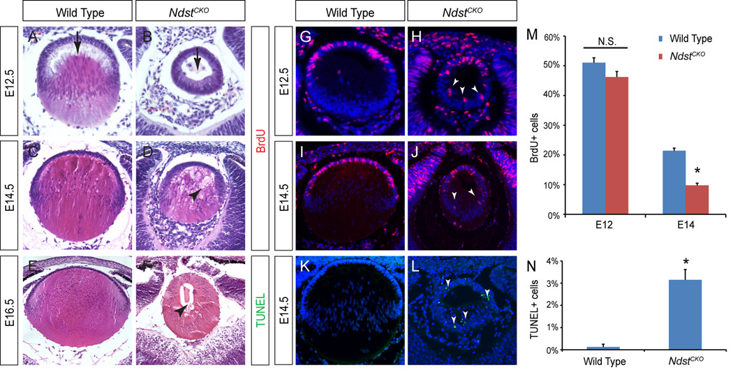
Figure 3. NdstCKO mutant lens phenotype.
(A–B) At E12.5, primary lens fiber cell elongation was delayed in NdstCKO mutant lenses (arrows). (C–D) E14.5 NdstCKO mutant lens fibers were disorganized amid numerous vacuoles (arrowhead). (E–F) Significant reduction in lens size and persistent lens vacuoles (arrowhead) were observed in the E16.5 NdstCKO mutant. At least ten embryos of each genotype were used for histology analysis. (G–N) Cell proliferation as shown by BrdU incorporation was significantly reduced in the NdstCKO mutants at E14.5 but not at E12.5, while cell death as shown by TUNEL staining was strongly elevated (Students t-test: N.S., not statistically significant; *P < 0.01; n=3).

