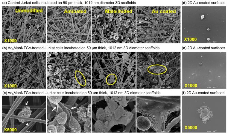
Figure 4.
Jurkat morphology when the cells are grown on different substrates with and without thiolated sialic acid display on their surfaces. Cells incubated with 3D scaffolds with the indicated surface chemistries in (a) the absence or (b) presence of Ac5ManNTGc at a magnification of ×1000 (x5000 for Ac5ManNTGc-treated cells is shown in (c)). Representative data for each of these conditions for 2D gold surfaces is shown in (d)-(f), respectively.

