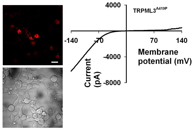
Fig. 5. TRP channels with constitutive activity show degeneration.
Left, Wide-field confocal images showing representative cell morphology of HEK cells transfected with the indicated construct. tdTomato-TRPML3A419P fluorescence (top) and DIC (bottom) images are displayed. Scale bar, 20 μm. Note that most fluorescent cells (top) also show degeneration, as displayed in transmitted image (bottom). Right, Representative I–V curve of whole-cell currents measured from HEK cells, expressing the indicated channel which displays robust inward rectifying currents.

