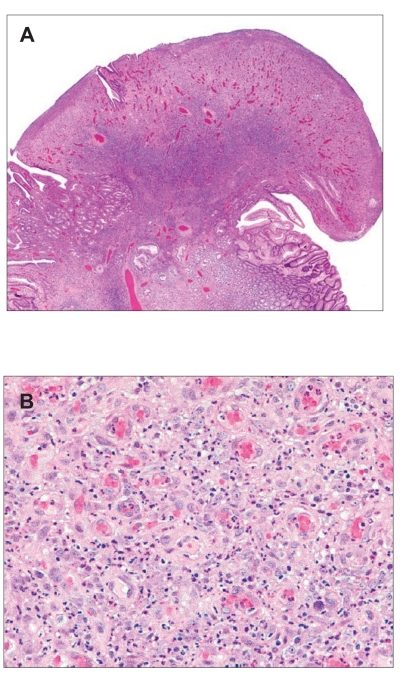
Figure 3.
Histology of pyogenic granuloma. Resection of the polyp at esophagogastroduodenoscopy revealed surface ulceration, acute inflammation, and lobular proliferation of capillary-sized vessels with a collarette of intact mucosa along the base. Figure 3B reveals details of the capillary structures, stromal edema, sparse lymphocytes, and marked neutrophilic infiltrate.

