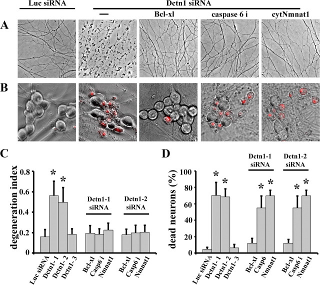
Figure 6.
Apoptotic pathways play important roles in axonal degeneration and cell death caused by Dctn1 knock down. A, Phase contrast microscopy showing axonal degeneration 7 d after DRG neurons were infected with lentiviruses expressing Luciferase (Luc) or Dctn1 siRNA. Extensive axonal degeneration occurred after Dctn1 knockdown that was blocked by Bcl-xl expression, caspase 6 inhibitor (caspase 6i) treatment, or cytNmnat1 expression. Axons of Luciferase siRNA-expressing neurons remained intact. B, Ethidium homodimer staining overlying phase contrast images of DRG neurons show extensive cell death in Dctn1 knockdown neurons compared with those expressing Luciferase siRNA. Bcl-xl expression blocked cell death after Dctn1 knockdown; however cytNmnat1 expression or caspase 6 inhibition failed to block neuronal death. C, Quantitative analysis of axonal degeneration (i.e., degeneration index) displayed in A demonstrated the protection afforded by Bcl-xl, caspase 6 inhibitor and cytNmnat1. D, Quantification of ethidium homodimer-positive neurons showed that only Bcl-xl expression blocks neuronal cell death caused by Dctn1 knockdown. The number of dead DRG neurons in each condition was scored and the percentage of dead neurons is displayed. Significantly different (*p < 0.001, Student's t test) from neurons expressing Luciferase siRNA.

